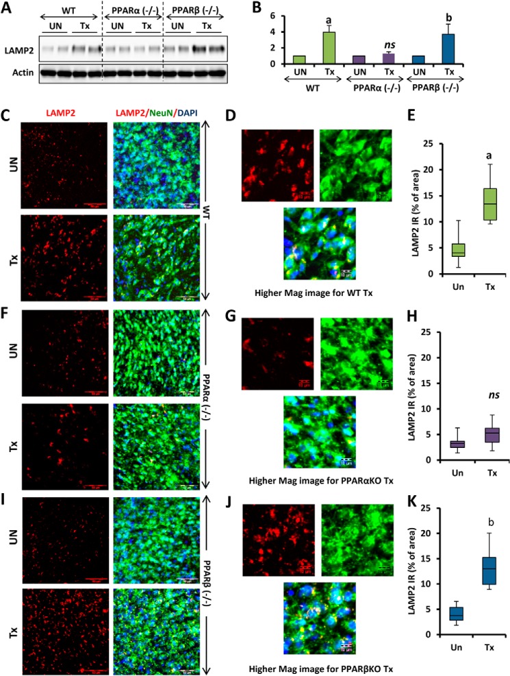
FIGURE 8.
Oral administration of gemfibrozil up-regulates LAMP2 in vivo in the cortex of WT and PPARβ−/−, but not PPARα−/−, mice. A, D, and G, WT, PPARα−/−, and PPARβ−/− mice (n = 6 in each group) were treated with 7. 5 mg/kg body weight/day gemfibrozil and 0.1 mg/kg body weight of all-trans retinoic acid (dissolved in 0.1% methylcellulose) or vehicle (0.1% methylcellulose) via gavage. After 60 days of treatment, mice were killed, and cortices from the animal brain were processed for immunoblot for LAMP2 (A). B, densitometric analysis of the immunoblot for LAMP2 (relative to β-actin). C, F, and I, cortical sections were double-labeled for LAMP2 (red) and NeuN (green). DAPI was used to visualize the nucleus. D, G, and J, higher magnification images showing localization of LAMP2 and NeuN in the cortical neuron of mice from the treatment group (WT, PPARα−/−, and PPARβ−/−). E, H, and K, quantification of LAMP2 immunoreactivity (LAMP2 IR) in untreated and treated samples from each group (WT, PPARα−/−, and PPARβ−/−) expressed as percentage of area. a, p < 0.05 versus WT control; b, p < 0.05 versus PPARβ−/− control; ns, not significant with respect to PPARα−/− control. At least 12 sections from each group (two sections per animal) were quantified using ImageJ. Scale bar, 50 and 10 μm (for higher magnification images). Tx, animals fed orally with 7.5 mg/kg body weight/day gemfibrozil and 0.1 mg/kg body weight day of all-trans-retinoic acid (dissolved in 0.1% methylcellulose); UN, animals fed orally with 0.1% methylcellulose (used as vehicle) (Un can be replaced with Veh).

