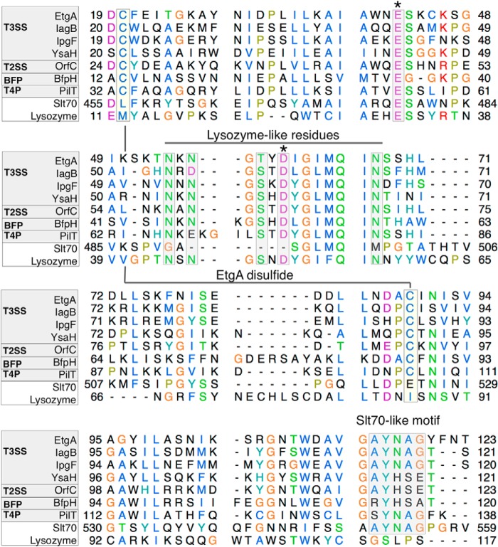
FIGURE 3.
Multiple sequence alignment of T3SS specialized PG-lytic enzyme EtgA with other macromolecular machine-associated peptidoglycan-cleaving enzymes, lytic transglycosylase Slt70 and C-lysozyme. Alignment of EtgA (E. coli, C7BUG6), IagB (Salmonella enterica serovar Typhi, P43018), IpgF (Shigella flexneri, Q07568), and YsaH (Yersinia enterocolitica, Q9KKJ1) with type II secretion system PG-lytic enzyme OrfC (Burkholderia pseudomallei, Q9ZF87), bundle-forming pilus PG-lytic enzyme Bfp H (E. coli 0127:H6, Q47073), type IVB pilus PG-lytic enzyme PilT (S. enterica serovar Typhi, Q9ZIU8), Slt70 lytic transglycosylase (E. coli, POAGC3), and lysozyme (Musca domestica, Q7YT16) is shown. The macromolecular machine-associated PG-lytic enzymes contain the glutamate general acid (denoted by *) present in both Slt70 and lysozyme, as well as an aspartate (denoted by *), which is present and required for catalysis in lysozyme but absent from Slt70. EtgA residues conserved with lysozyme that form a hydrogen-bonding network with the conserved Asp are boxed and labeled as Lysozyme-like residues. Cysteines that form a disulfide bond in EtgA and are conserved with other T3SS, type IV pili, and bundle-forming pili, and the type II secretion system PG-lytic enzymes are boxed and labeled. A motif conserved between Slt70 and T3SS-associated PG-lytic enzymes is also boxed and labeled. Sequence alignment was done using clustal omega and the image was generated using Chimera. Swiss-Prot/TrEMBL accession numbers for each sequence are shown above in brackets.

