FIGURE 5.
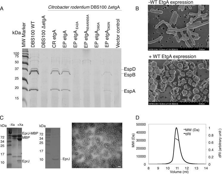
Analysis of EtgA activity in vivo and interaction with the inner rod. A, type III secretion assay of C. rodentium etgA mutant complemented with EPEC etgA and its site-directed mutants. C. rodentium DBS100 ΔetgA was complemented with EPEC etgA with mutations of key conserved active site residues. T3SS secreted proteins (translocon components EspB and EspD, as well as EspA filament) from C. rodentium grown in DMEM were analyzed by 16% SDS-PAGE and stained by Coomassie G250. B, scanning electron microscopy images of BL21 (DE3) E. coli without expression of EtgA (top panel) and after induction of EtgA expression (bottom panel). EtgA was fused to a PelB signal peptide for export to the periplasm. D, stoichiometry of EtgA and inner rod (EscI) complex by size exclusion chromatography-multiangle light scattering. EscI (10–137) and EtgA (19–152) were co-purified, injected over a Superdex 75 10/300 column, and analyzed by multiangle light scattering. EscI (10–137) and EtgA (19–152) were shown to form a monodisperse complex of 31,400 Da (± 0.4%), corresponding to a 1:1 ratio. C, purification of MBP tagged EprJ (EscI homolog in EHEC) by amylose affinity chromatography (left gel) followed by cleavage of the MBP tag and size exclusion chromatography (right gel). Purified EprJ was imaged by negative stain electron microscopy. The white scale bar corresponds to 100 nm. MW, molecular mass.

