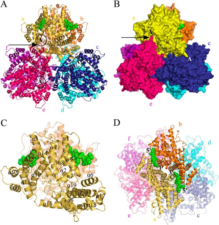
FIGURE 2.
Views of the Dgt hexameric crystal structure. A, top view of the hexameric organization showing Dgt as a stack of two trimers. The top trimer consists of monomers labeled a (yellow), c (blue), and e (hot pink). The bottom three monomers are labeled b (orange), d (cyan), and f (magenta). The two DNA molecules (modeled as a CCC trinucleotide) bound at the interface of monomers a and b are shown as solid green spheres. The active site region of monomers a, c, and e is indicated with a circled capital A. The arrow is pointing toward a small loop in monomer e (in hot pink) that reaches out into the active site of monomer a (in yellow). See text for details. B, surface representation of structure as shown in panel A. The putative active sites of top monomers (a, c, and e) are indicated with capital A. The long arrow points toward one example of a small loop in each monomer that reaches out into the active site of the next clockwise monomer; see text and Fig. 4D for details. C, ribbon diagram of monomer a (in yellow) with individual helices numbered α1 to α21. The two DNA molecules are in green. D, side view of the Dgt hexamer with the bound DNA (green) at the interface of a and b. The 5′ → 3′ orientations of the DNA are also indicated.

