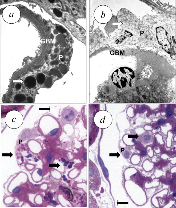
Fig. 4.

Effect of 4 weeks of treatment with the ERA darusentan on renal structure in established FSGS [39]. (a) Untreated animal, transmission electron microscopy demonstrates GBM hypertrophy and podocyte injury with diffuse foot process effacement and vacuolar degeneration involving autophagy [113]. (b) ERA-treated animal, showing regression of GBM hypertrophy and disappearance of podocyte vacuoles. (c) Untreated animal, light microscopy image (haematoxilin/eosin) demonstrating hypertrophy of podocytes with enlarged nuclei, prominent nucleoles and vacuolar degeneration due to autophagy. (d) Treated animal, showing normal sized podocyte nuclei and virtually complete disappearance of vacuolar degeneration (arrows). Scale bar, 10 μm (c, d). Panels adapted from reference [39] and reproduced with permission of the publisher.
