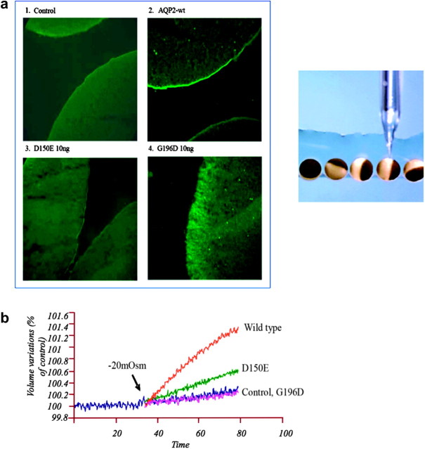
Fig. 1.
(a) Immunofluorescence of AQP2 expressed in oocytes. Oocytes were not injected (1 control) or injected with either AQP2-wt (2, 1 ng), AQP2-D150E (3, 10 ng) or AQP2-G196D (4, 10 ng) messenger RNAs and incubated for 3 days prior to assay. Oocytes were immunostained and visualized with antibodies to AQP2. The injection process is represented in the right of the figure [21]. (b) Determination of water permeabilities (Pf) of wild-type (WT) AQP2 and mutants expressed in Xenopus oocytes. Oocytes were injected with either AQP2-wt (1 ng), AQP2-D150E (10 ng) or AQP2-G196D (10 ng) messenger RNAs and incubated for 2 days prior to assay. Determination of water permeabilities was performed by evaluation of volume increase in oocytes as induced by a 20-mosmol/kg H2O hypotonic shock [21].

