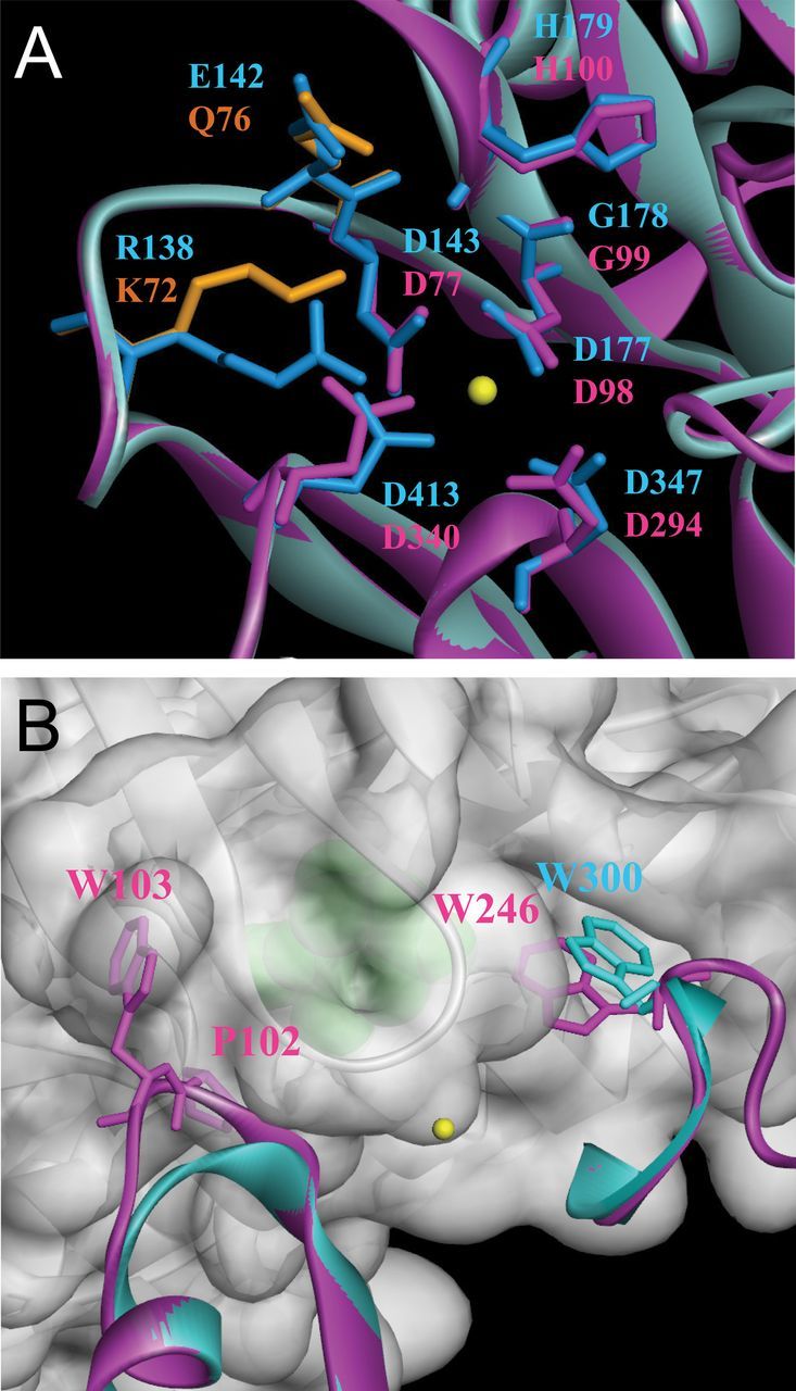
Fig. 3.

Homology-based structural modelling of AtPP2CF1. Ribbon representation of AtPP2CF1. (A) Close-up view of the putative active site of AtPP2CF1. The key residues comprising the active site of ABI1 are shown as blue-stick representations. Among the active site residues of AtPP2CF1, residues identical to ABI1 are shown as magenta-stick representations, and those specific for AtPP2CF1 are depicted as orange-stick representations. Note that at least one metal ion (yellow sphere) was bound to the active site of AtPP2CF1. (B) Close-up view of the AtPP2CF1 regions corresponding to the primary-binding interface of ABI1 with PYL1. PYL1 (grey) is shown in a semi-transparent surface representation. (+)-ABA is shown as a green sphere. For simplicity, only two regions of AtPP2CF1 (magenta), which correspond to the β3-α1 loop and the β9-β10 loop of ABI1 (blue), are shown. Trp246 of AtPP2CF1, Trp300 of ABI1, and Pro102 and Trp103 of AtPP2CF1, which correspond to an insertion of two amino acids in the β3-α1 loop of consensus motif 4, are highlighted in the stick representation.
