Fig. 3.
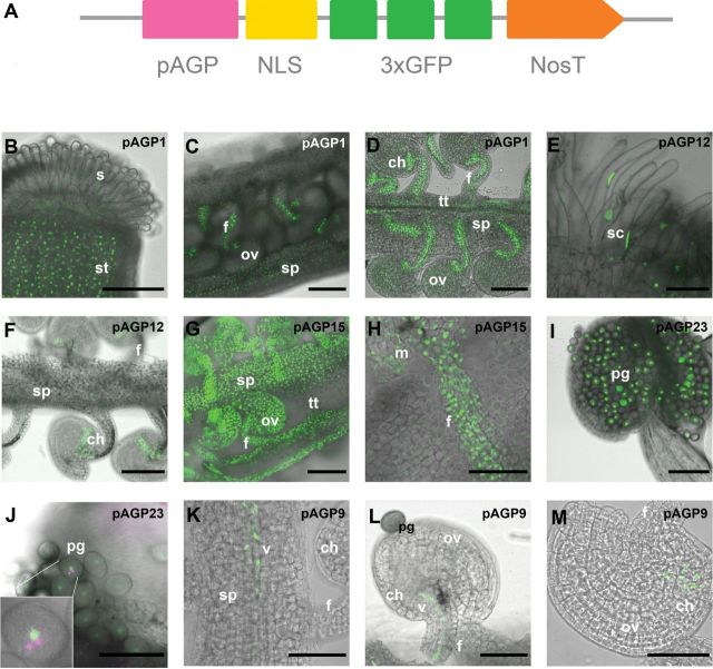
Schematic representation of the expression cassette used in this study, and the resulting GFP signal shown in Arabidopsis reproductive tissues. (A) Expression cassette showing the relative position of the promoter sequence (pAGP), NLS, a fusion of three GFPs (3×GFP), and the terminator Nos (NosT). (B–D) NLS:3GFP expression driven by the AGP1 promoter in the style tissues (B), the opened pistil, and the funiculus and septum tissues (C), and seen in more detail in the transmitting tissue, funiculus, and the chalazal pole of the ovule (D). (E–F) NLS:3GFP expression under the control of the AGP12 promoter was observed in the stigmatic cells (E) and in the chalazal pole of the ovule (F). (G–H) NLS:3GFP expression driven by the AGP15 promoter was detected in the ovule integuments, funiculus, and septum, but absent from the transmitting tissue (G). In (H) the GFP signal is seen in more detail in the nuclei of the funiculus. (I, J) NLS:3GFP under the control of the AGP23 promoter is absent in all the sporophytic tissues (I), with its expression restricted to the pollen grain, and, as can be seen in the insert in (J), DAPI staining (here in magenta) revealed this expression to be limited to the vegetative cell of the pollen grain; DAPI-stained germinative nuclei are visible (white arrowheads). (K–M) NLS:3GFP signals expressed by the AGP9 promoter. Signals were observed in the vascular bundle of the transmitting tract (K) and funiculus (L) as well as in the chalazal pole of the ovule (M). All the flowers used in these observations were at stages 12 and 13 according to Smyth et al. (1990). ch, Chalaza; f, funiculus; m, micropyle region of the ovule; ov, ovule; pg, pollen grain; s, stigma; sc, stigmatic cell; sp, septum; st, style; tt, transmitting tract; v, vascultature. Bars, 100 μm (B–G, I); 50 μm (H, K–M); 20 μm (J).

