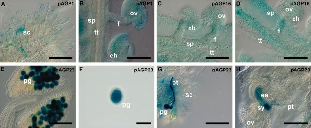
Fig. 4.

Histochemical localization of GUS activity in transgenic Arabidopsis reproductive tissues expressing the pAGP:GUS fusion genes. (A, B) GUS activity driven by the AGP1 promoter is detected in the stigmatic cells (A) and the transmitting tract, funiculus, and integument cells (B). (C, D) GUS activity driven by the AGP15 promoter observed in the ovule integuments, funiculus, and septum cells. (E–H) Strong GUS activity driven by the AGP23 promoter was identified inside pollen grains (E) and (F) and the growing pollen tube (G). Upon fertilization, inside the embryo sac, a strong staining is observed at the local where the pollen tube bursts (H), followed by a weak staining that spread inside the whole embryo sac (H). Flowers of stages 12 and 13 (Smyth et al., 1990) were used in this study. ch, Chalaza; es, embryo sac; f, funiculus; ov, ovule; pg, pollen grain; pt, pollen tube; sc, stigmatic cell; sp, septum; sy, synergid; tt, transmitting tract. Bars, 100 μm (A–E, G, H); 50 μm (F).
