Fig. 5.
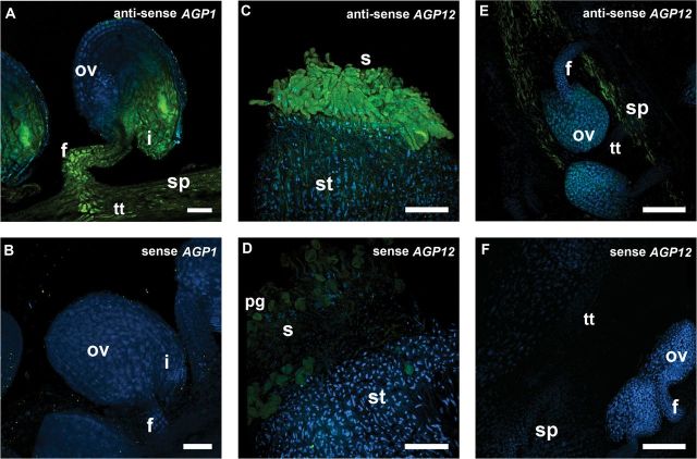
FISH localization of AGP1 and AGP12 transcripts in Arabidopsis pistil tissues. Merged images of FISH signals (green) and DAPI staining of nuclei (blue) are shown. (A) AGP1 transcripts were detected in the funiculus, transmitting tissue, and integuments. (C, E) AGP12 transcripts were localized in the stigmatic cells and along the septum tissues. (B, D, F) FISH controls with the sense probe for AGP1 in ovules (B) and for AGP12 in stigma (D) and ovules (F). All the flowers used in these observations were at stages 12 and 13 according to Smyth et al. (1990). f, Funiculus; i, integuments; pg, pollen grain; ov, ovule; s, stigma; sp, septum; st, style. Bars, 25 μm (A, B); 75 μm (C–F).

