
Fig. 6.
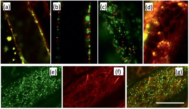
Protein–GFP localization. Transient co-localization analysis of AtPRPL1–GFP and RFP-tagged markers for the (a) plasma membrane, (b) Golgi apparatus, (c) peroxisomes, and (d) ER. (e) AtPRPL–GFP, (f) mCherry-fused ER marker ER-rb CD3-960, and (g) overlay of both in 35S::AtPRPL1-GFP lines stably transformed with the ER marker. Scale bar=10 μm.