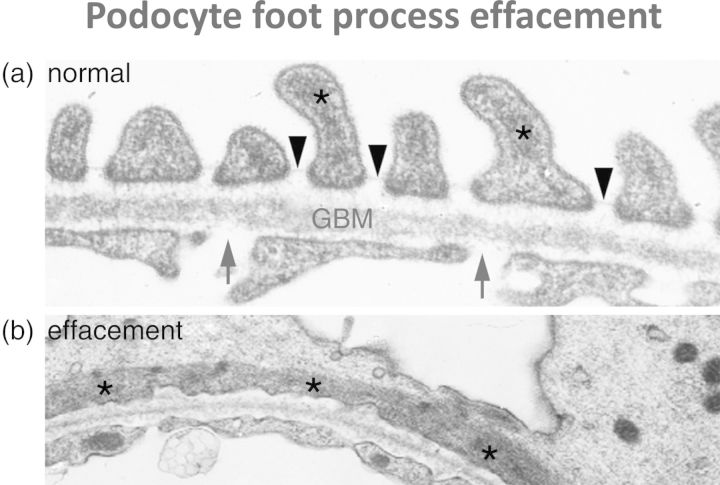
Fig. 2.
Transmission electron micrographs of the human glomerular capillary wall. (a) shows the normal appearance: arrows indicate endothelial fenestrations, arrowheads indicate filtration slits between podocyte foot processes and asterisks indicate actin filaments in podocyte cytoplasm. Note the actin filaments focused in podocyte foot processes. (b) shows podocyte foot process effacement as seen in proteinuric states: note flattening of the actin filaments (asterisked) longitudinally associated with the loss of normal foot process architecture.

