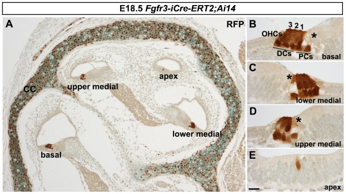
Fig. 1. Recombination pattern obtained with the Fgfr3-iCre-ERT2 mice.
(A–E) Fgfr3-iCre-ERT2;Ai14(tdTomato) mice treated with tamoxifen at E13.5 and E14.5 show recombination in the organ of Corti and in the cochlear capsule, revealed by red fluorescence protein (RFP) immunohistochemistry on paraffin sections at E18.5. Recombination follows a base-to-apex gradient along the length of the cochlear duct. All Deiters' and pillar cells and OHCs in the basal (B) and lower medial coils (C) are recombined. A large part of these cells are recombined in the upper medial coil (D), but only rare recombined cells are found in the apex (E). Inner hair cells (asterisks in B–D) are not stained for RFP. The three OHC rows are numbered. Abbreviations: OHCs, outer hair cells; PCs, pillar cells; DCs, Deiters' cells; CC, cochlear capsule. Scale bar shown in E: A, 25 µm; B–E, 7 µm.

