
Fig. 2. Cdc42 in situ hybridization and immunostaining in the organ of Corti at E18.5.
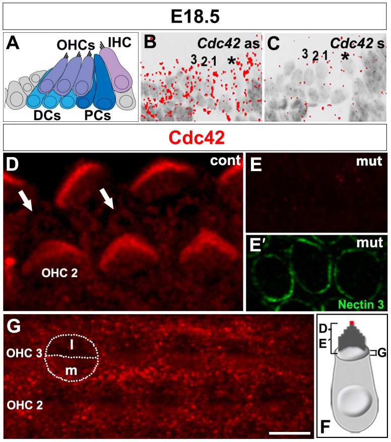
(A) Schematic representation of the cytoarchitecture of the organ of Corti at E18.5. (B,C) The antisense (as) probe shows ubiquitous Cdc42 mRNA expression in the sensory epithelium (B). The Cdc42 sense (s) probe reveals the background level. Asterisk marks IHC. The three OHC rows are numbered. (C). (D–F) Confocal images of cochlear whole mount specimens stained for antibodies against Cdc42. Strong expression is seen in the stereocilia of hair bundles and weaker expression around centrioles (arrows), lateral to the bundle (D). Double-labeling shows the absence of Cdc42 immunostaining in the Cdc42loxP/loxP;Fgfr3-iCre-ERT2 mice (E), while nectin 3 marks the junctions between OHCs and Deiters' cells (E′). Control animals show weak Cdc42 staining in the medial surface domain of OHCs. Contours of an OHC are outlined and a line is drawn to separate the medial (m) and lateral (l) surface domains. Staining is also found at the level of adherens junctions between OHCs and Deiters' cells (G). (F) Schematic picture shows the planes and areas covered in the immunofluorescence views. The red dot marks the site of the kinocilium and basal body. Abbreviations: IHC, inner hair cell; OHCs, outer hair cells; DCs, Deiters' cells; PCs, pillar cells. Scale bar shown in G: A–C, 20 µm; D, 3 µm; E–G, 5 µm.