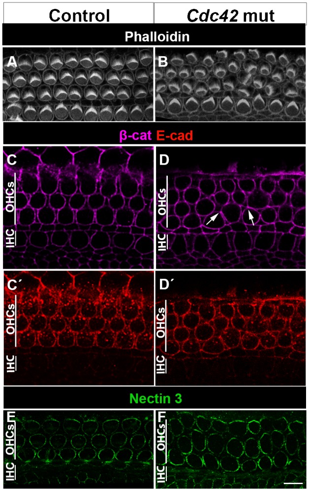
Fig. 3. Cellular disorganization, but unaltered expression of adhesion proteins in the organ of Corti of Cdc42loxP/loxP;Fgfr3-iCre-ERT2 mice at E18.5.

(A,B) Phalloidin labeling shows the organization of OHCs into three rows in the control specimen. In the organ of Corti of the mutant mouse, misplacement of some OHCs in between the rows is seen. The organization of the row of unrecombined IHCs is unaltered. Both views are from the medial coil. (C–D′) Confocal views at the level of adherens junctions show comparable expression of β-catenin (C,D) and E-cadherin (C′,D′) in the two genotypes. Arrows in D point to atypical intercellular contacts. (E,F) Nectin 3 immunofluorescence is comparable in the contact sites between OHCs and Deiters' cells in both genotypes. Abbreviations: IHC, inner hair cell; OHC, outer hair cell; β-cat, β-catenin; E-cad, E-cadherin. Scale bar shown in F: A,B, 6 µm; C–F, 5 µm.
