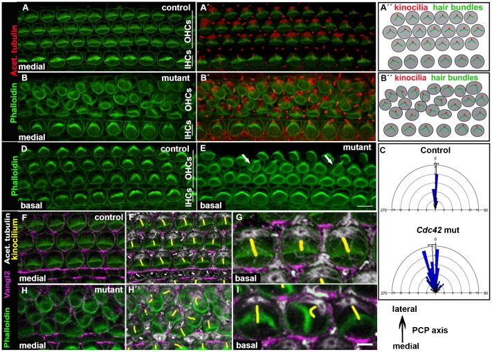
Fig. 5. Misorientation of stereociliary bundles of outer hair cells of Cdc42loxP/loxP;Fgfr3-iCre-ERT2 mice.
Confocal images of whole mount specimens at E18.5. (A,A′) Shown by double-labeling, the medial coil of the control cochlea displays uniform orientation of phalloidin-labeled hair bundles and their laterally pointing vertices. The kinocilia, positive for acetylated tubulin, are located at vertices. (B,B′) The medial coil of the mutant cochlea shows several OHCs with misoriented bundles. (A″,B″) Schematic representations of hair bundle orientations. (C) Quantification (see materials and methods) shows the distribution of randomly oriented bundles in the medial coil of mutant cochleas. (D) Phalloidin labeling shows normal hair bundle orientation in the basal coil of the control cochlea. (E) The basal coil of the mutant specimen shows occasional OHCs with misoriented bundles (arrows), but hair cell rows are normally organized. (F–G) Triple-labeling for F-actin, Vangl2 and acetylated tubulin (kinocilia pseudocolored in yellow) shows Vangl2 expression in the contact sites between the OHC's medial wall and Deiters' cells in the control specimen. (H,H) In the medial coil of the mutant cochlea, Vangl2 expression pattern is influenced by cellular disorganization. (I) In the basal coil of the mutant cochlea where cellular disorganization is absent, Vangl2 expression domain is similar as in the control specimen (compare to Fig. 5G). Abbreviations: OHC, outer hair cell; IHC, inner hair cell. Scale bar shown in I: A–E, 6 µm; F–H′, 5 µm; G,I, 2 µm.

