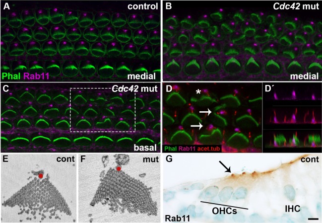
Fig. 6. Disturbances in stereociliary bundle morphology, but maintained Rab11a expression in outer hair cells of Cdc42loxP/loxP;Fgfr3-iCre-ERT2 mice.
(A) Confocal view of a whole mount specimen from the medial part of the control cochlea at E18.5 shows Rab11 expression close to the vertices of phalloidin-labeled hair bundles. (B,C) Both in the medial and basal coil of the mutant cochlea, Rab11 is expressed similarly as in the control specimen. (D,D′) Higher magnification view of the boxed area in C shows partial colocalization of acetylated tubulin, a kinocilium marker, and Rab11 at the base of kinocilia. This colocalization is best revealed in transverse plane (D′). A part of OHCs in this mutant specimen show dysmorphic bundles accompanied by off-centered kinocilia (arrows). Note that bundle dysmorphology exists also in normally oriented OHCs (asterisk). (E,F) SBEM images of OHC stereociliary bundles of control and mutant cochleas at E18.5. In the control specimen, kinocilium (red dot) is located at the vertex of the V-shaped hair bundle. In the mutant specimen, kinocilium is misplaced and the bundle is dysmorphic, with no clear vertex. (G) Also transverse sections immunohistochemically stained for Rab11 demonstrate expression in the apex of OHCs. Abbreviations: OHC, outer hair cell; IHC, inner hair cell. Scale bar shown in G: A–D″, G, 5 µm; E,F, 1 µm.

