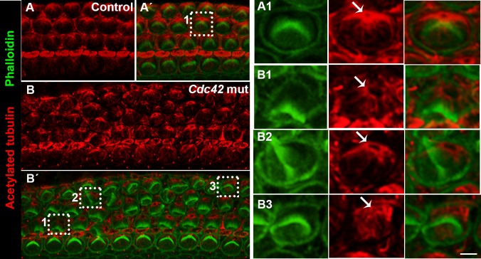
Fig. 7. Disorganized apical microtubule network in outer hair cells of Cdc42loxP/loxP;Fgfr3-iCre-ERT2 mice.
Confocal views of cochlear whole mount specimens dissected from the medial coil of control and mutant mice at E18.5. (A–B′) Specimens double-labeled for acetylated tubulin, marking microtubules and basal body, and for phalloidin that labels the hair bundle. Z-stacks cover the level from the basal body to the apicalmost astral microtubules of OHCs. Boxed OHCs in (A′,B′) are shown in higher magnification in A1,B1,B2,B3. (A1) Control specimen shows the localization of the basal body (arrow) close to the vertex of hair bundle and ordered radiation of microtubules at the OHC surface. (B1–B3) In the mutant specimen, the microtubule network is rotated with respect to hair bundle misorientation and microtubules around the basal body are disorganized (B1). Some OHCs show complete loss of the ordered microtubular radiation (B3). Scale bar shown in B3: A–B′, 5 µm; A1–B3, 2 µm.

