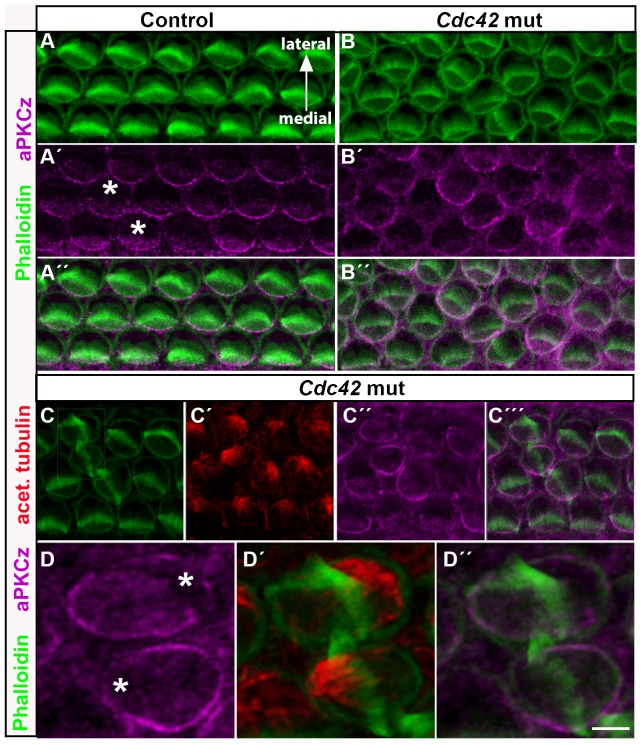
Fig. 8. Altered aPKCζ expression in outer hair cells of Cdc42loxP/loxP;Fgfr3-iCre-ERT2 mice.
Confocal images of whole mount specimens dissected from the medial cochlear coil of control and mutant mice at E18.5 and labeled for F-actin, acetylated tubulin and aPKCζ. All images are shown in medial-lateral orientation, as indicated in (A). (A–A″) In the control specimen, aPKC is expressed in the cortical and, weaker, in the cytoplasmic domain at the OHC surface, medial to the hair bundle. Asterisks mark the lateral, aPKC-negative domain. (B–B″) In the mutant specimen, both cortical and cytoplasmic aPKC expression domains are rotated with respect to hair bundle misorientation. (C–C′″) In the mutant specimen, triple-labeling for acetylated tubulin, phalloidin and aPKC shows that the rotation of aPKC expression parallels the rotation of the astral microtubule network and hair bundle misorientation. (D–D″) High magnification views of two OHCs with abnormal bundles. The OHC below has a completely turned bundle. Asterisks mark the concise aPKC-free area in these cells (compare to control OHCs in Fig. 8A′). Scale bar shown in D″: A–C′″, 5 µm; D–D″, 2 µm.

