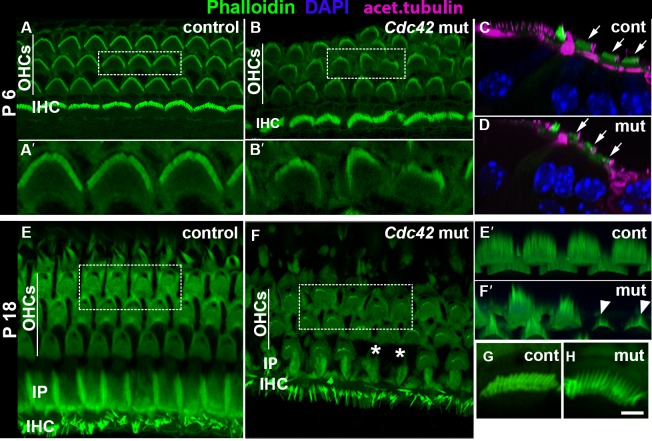
Fig. 9. Impaired stereociliogenesis in outer hair cells of Cdc42loxP/loxP;Fgfr3-iCre-ERT2 mice postnatally.
Confocal views of whole mount specimens from the medial coil of the cochlea. Boxes in A,B,E,F are enlarged in A',B',E',F', respectively. (A,A′) Phalloidin labeling shows the typical hair bundle morphology in the control specimen at P6. (B,B′) In the mutant cochlea at P6, all OHCs with stereociliary bundles are present, but some bundles are misoriented and have a fragmented or stunted appearance. (C,D) Degeneration of OHC bundles is also seen in transverse views where DAPI labels nuclei and acetylated tubulin marks kinocilia and microtubules. Arrows point to outer hair cells. (E,E′) Phalloidin labeling reveals the normal cellular organization and hair bundle morphology in the control specimen at P18. (F,F′) In the mutant cochlea, scattered OHC loss is seen (asterisks in F). Also, OHC bundles are fragmented and often composed of very short stereocilia (arrowheads in F′). (G,H) Unrecombined IHCs of mutants show stereociliary bundles comparable to controls. Abbreviations: OHCs, outer hair cells; IHC, inner hair cell; IP, inner pillar cell. Scale bar shown in H: A–D, 5 µm; E′,F′ 3 µm; A′,B′,G,H, 2 µm.

