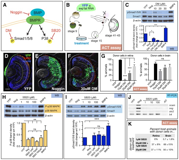
Fig. 1. Repression of canonical and/or non-canonical BMP signaling fails to replicate the retina-inducing efficiency of Noggin.
(A) Schematic of the canonical and non-canonical BMP pathways and the downstream signaling molecules, Smad1/5/8 and p38 MAPK, respectively. Small molecule inhibitors dorsomorphin (DM) and SB203580 (SB20) were used to specifically inhibit canonical and non-canonical signaling, respectively. (B) Diagram of experimental design for animal cap transplant (ACT) assay. YFP RNA with and without experimental (exp'tal) RNA was injected into both cells of a two-cell stage embryo. The animal cap was removed from the blastula (stage 9) and cultured until sibling embryos formed a neural plate (stage 15). Part of the animal cap was then transplanted into the eye field of a host embryo, which was grown until the eye differentiated (stages 41 to 43). Cryostat sections were analyzed for the presence of YFP+ transplanted cells. (C–G) Analysis of canonical signaling pathway. (C) Western blots were used to detect pSmad1/5/8, Smad1, and β-actin in stage 15 animal caps treated with DM. Treatment with 20 and 30 µM of DM is sufficient to suppress pSmad1/5/8 as efficiently as Noggin (Nog). (D–F) Representative images of transplanted cells in the retina. Treating animal caps with 30 µM of DM drives retinal specification in only 75% of embryos. Scale bars, 50 µm. Dashed lines lie on outer and inner plexiform layers, separating the three retinal layers. (G) The number of animals with transplanted cells in the eye were identified by scoring cryostat sections stained for YFP (green), rod photoreceptor marker, XAP2 (red), and DAPI (blue). Quantification of retinal integration efficiency, depicted as % of animals with YFP+ donor cells in the retina or brain. YFP, n = 44, Nog, n = 90; 10 µM DM, n = 46; 20 µM DM, n = 154; 30 µM DM, n = 73. (H–K) Analysis of non-canonical BMP pathway. (H) Western blot analysis of animal caps treated with SB203580 (SB20). As expected, activity of p38 (P-p38) is inhibited in caps treated with Noggin and SB20. (I) Canonical signaling through pSmad1/5/8 is not affected by SB20 treatment. (J) SB20 treatment fails to induce the expression of neural genes, ncam, nog, and otx2, compared to DNA histone H4 (h4) loading control; N = 3. (K) Animal caps treated with 1 µM SB20 fail to incorporate into host retina, but a few animals have transplanted cells in the brain (n = 53). Treatment with 1 µM SB20 and 20 µM DM, compared to 20 µM DM alone, produces the same percentage of host animals with transplanted cells in the retina, while there is a slight increase the number of animals with donor cells in the brain (n = 97). Western blots, WB; reverse transcription-PCR, RT-PCR; animal cap transplant assay, ACT assay. Error bars = ±s.e.m.; *p<0.05; **p<0.01; ***p<0.001; ns, not significant.

