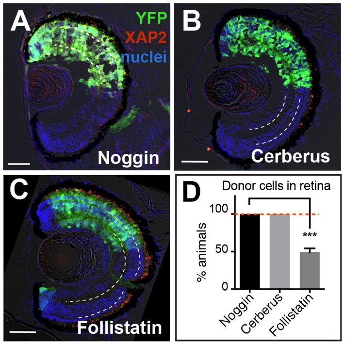
Fig. 7. Follistatin is less efficient than Cerberus or Noggin at specifying retina.
(A–C) YFP+ donor cells expressing Noggin, Cerberus, and Follistatin all contribute to the retina. However, Noggin- and Cerberus-treated cells formed retina in all animals while cells expressing Follistatin had a significantly lower retinal integration efficiency (D). On retinal sections, dashed white lines lie on outer and inner plexiform layers. Green, YFP donor cells; red, rod photoreceptor marker XAP2; blue, DAPI staining. YFP, 500 pg, n = 29; Nog, 20 pg, n = 65; Cerberus, 1.6 ng, n = 77; Follistatin, 1200 pg, n = 53; N = 3. Scale bars, 50 µm. Error bars = ±s.e.m.; ***p<0.001. Dashed red line on the graph marks Noggin/Cerberus treatment, highlighting the difference from Follistatin treatment.

