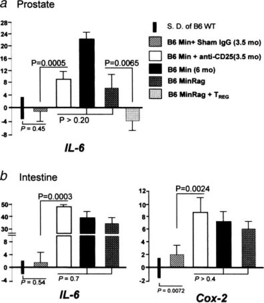
Figure 4.
mRNA expression levels of IL-6 and Cox-2 in the murine prostate and bowel. There were significant increases in expression of IL-6 in (a) prostate and (b) ileum of Min mice treated with anti-CD25 antibody, and also in Rag-deficient Min mice, when compared to sham-treated Min counterparts. Supplementation with TREG cells from wt donor mice decreased expression of IL-6 in prostate tissue, whereas depletion of CD25+ cells increased IL-6 gene expression in prostate tissue. Assay used tissues from 5 to 8 mice per treatment group. For comparison of mRNA levels, the target mRNA was normalized to that of the housekeeping gene GAPDH. Numbers on the y-axis represent mean fold change of target mRNA levels in reference to the control levels (B6 wt, defined as 0, standard deviation represented by solid bars). mos, age in months upon necropsy.

