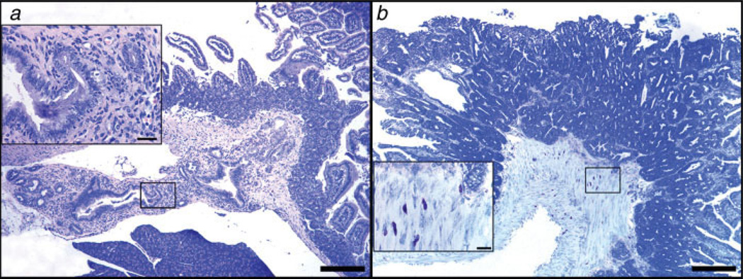
Figure 6.
Depletion of lymphocytes correlates with an invasive neoplastic phenotype in the small intestine of Apcmin/+mice. (a) Ampullary cancer arising from pancreatic duct epithelium in 25% (2/8) of 3-month-old ApcMin/+ mice after undergoing depletion of CD25+ cells (N = 8 mice per trial). High magnification (inset in a) reveals features of the abnormal pancreatic duct epithelium including pseudostratification and nuclear pleomorphism with associated inflammation. (b) Ileum of Rag2−/− ApcMin/+ mouse at 6 months of age revealing submucosal and incipient tunica muscularis invasion of neoplastic glands in an adenomatous polyp. Large numbers of mast cells infiltrate the submucosa and muscle layers at the base of the polyp. The higher magnification (inset) reveals the close topographic association of mast cells with the invasive front of the tumor. (a) Hematoxylin and Eosin. (b) Toluidine Blue. Bars a and b: 250 µm; insets: 25 µm. [Color figure can be viewed in the online issue, which is available at www.interscience.wiley.com.]

