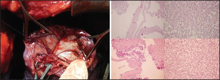
Figure 1.

The left panel show operative view of the opened hydatid cyst cavity, and the right panel show histopathological examination showing typical Aspergillus hyphae and membrane of the hydatid cyst (H and E, ×40) and Aspergillus hyphae makes a 45° angle on the fungus ball (H and E, ×400)
