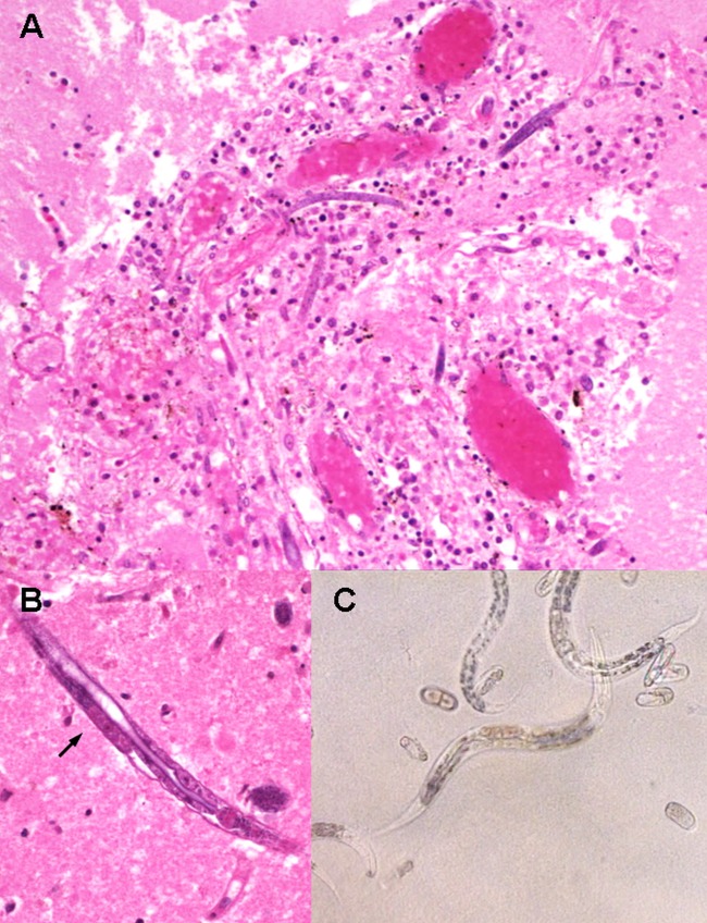
FIG 2.
(A) Hematoxylin and eosin (H&E) stain of brain tissue (under ×100 magnification) demonstrates perivascular inflammation with predominant macrophages and lymphocytes surrounding H. gingivalis larvae. (B) Third-stage larvae stained with H&E under ×400 magnification show presence of premature genital primordium (black arrow) and bulb of esophagus (at right end of nematode). (C) Microscopy examination of agar plate culture (×400 magnification) shows H. gingivalis larvae and eggs in various stages of development.

