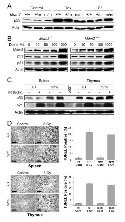
Figure 3. Mdm2C305F mice retain intact p53 response to DNA damage.
(A) Mdm2+/+, Mdm2m/+ and Mdm2m/m MEFs were mock treated (Control) or treated with 1 μM doxorubicin (Dox) or 50 J/m2 of ultraviolet C (UV). The cells were harvested 18 hours post treatment and immunoblotted for p53 and Actin.
(B) Mdm2+/+ and Mdm2m/m MEFs were treated with increasing dosage of Dox for 18 hrs. Cell extracts were analyzed for p53, p21 and Mdm2.
(C) Induction of p21 in spleen and thymus after γ-irradiation. Spleen and thymus were harvested from Mdm2+/+ and Mdm2m/m mice 18 hours after 8-Gy whole body γ-irradiation. Protein extracts were assayed for protein levels of p53, p21, and Actin. Spleen protein extract from a p53−/− mouse was used as a negative control.
(D) Apoptotic response to whole body γ-irradiation. The level of apoptosis in spleen and thymus was determined by TUNEL assay, carried out on paraffin embedded tissues from mice 18 hours after receiving 8-Gy of whole body γ-irradiation. The percentage of TUNEL positive cells was quantified and averaged from three identically treated mice. Scale bars represent 75 μm. Error bars in all cases represent ± SD.

