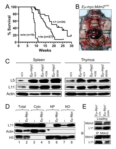
Figure 5. Myc-induced lymphomagenesis is accelerated by Mdm2C305F mutation.
(A) Survival of Eμ-Myc transgenic mice. Kaplan-Mierer survival curve shows a median survival of 20, 15, and 9 weeks for Eμ-Myc;Mdm2+/+ (+/+, n= 24), Eμ-Myc;Mdm2+/m (+/m, n= 27) and Eμ-Myc;Mdm2m/m (m/m, n= 18) mice, respectively.
(B) Lymphoma onset in Eμ-Myc;Mdm2m/m mice. Image shows dissection of a 58-day old Eμ-Myc;Mdm2m/m mouse with typical enlargement of liver, spleen, thymus and lymph nodes, including enlargement of Peyer’s patches and mesenteric lymph nodes.
(C) Extracts from spleen and thymus of wild type (+/+) and tumor bearing Eμ-Myc transgenic mice were analyzed for L5 and L11.
(D) Accumulation of L11 in nucleus in Eμ-Myc transgenic mouse splenocytes. Lysates of splenocytes from wild type and non-tumor bearing Eμ-Myc transgenic mice were subjected to fractionation as described in Experimental Procedures. The total (Total), cytoplasmic (Cyto), nucleoplasmic (NP), and nucleolar (NO) fractions were isolated and analyzed for L11. Actin (cytoplasmic) and Histone H3 (nuclear) were used as fractionation markers.
(E) L11-Mdm2 binding in Eμ-Myc transgenic splenocytes. Lysates from spleens of 4-week-old, non-tumor bearing mice were immunoprecipitated with anti-Mdm2 antibody and immunoblotted as indicated. Loading represents 5% of total lysate used for IP.

