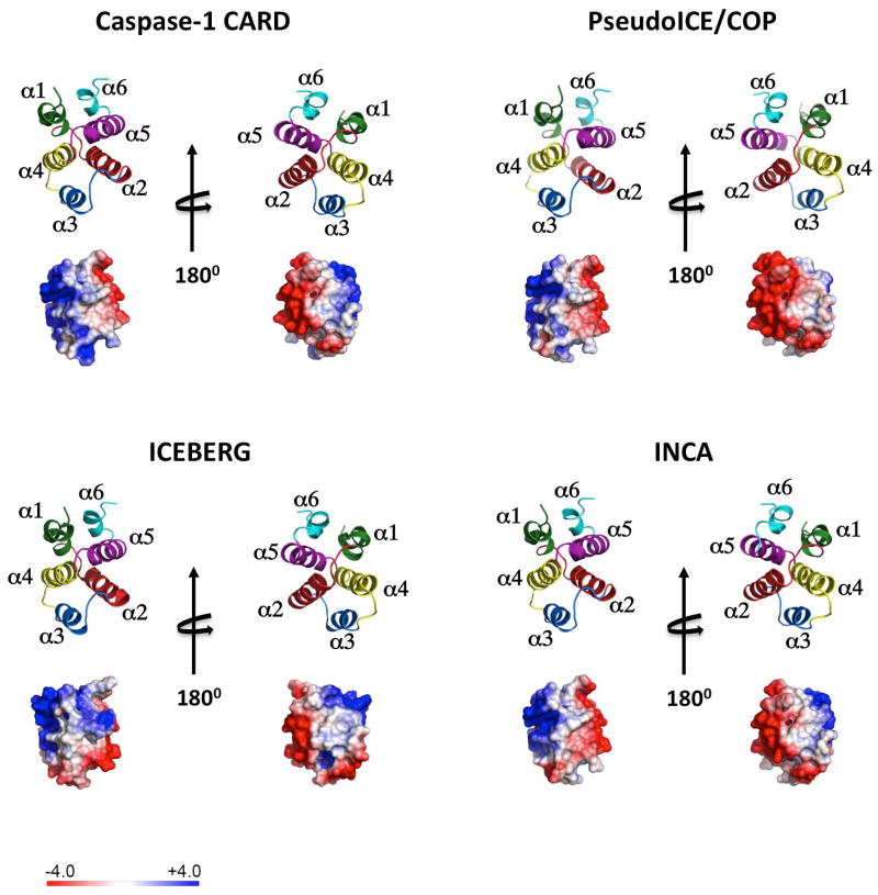
Fig. 3. The structure of the caspase-1CARD, Pseudo-ICE/COP, INCA, and ICEBERG is shown as ribbon diagram and the electrostatic surface is displayed on a scale of -4 kT/e (red) to 4kT/e (blue).
The structure of the ICEBERG (1DGN) (83) has been resolved. Homology modeling (SWISS-MODEL) (65) is shown for the caspase-1CARD, Pseudo-ICE/COP, and INCA based on ICEBERG. Helices are colored in green (α1), red (α2), blue (α3), yellow (α4), purple (α5) and turquoise (α6).

