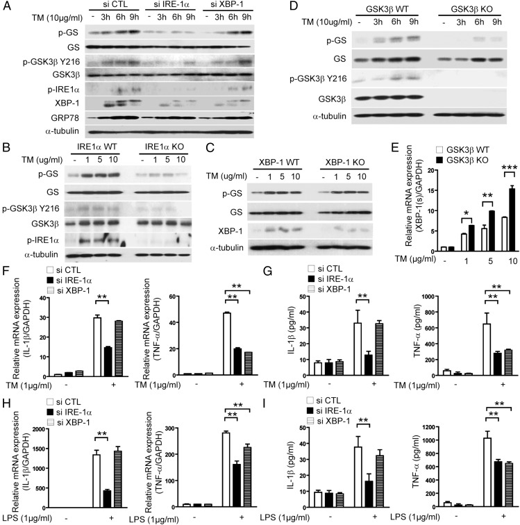
FIGURE 2.
ER stress–induced IRE1α activation regulates differential proinflammatory cytokine gene expression via activation of GSK-3β and XBP-1. (A) RAW 264.7 cells were transfected with specific siRNA against IRE1α, XBP-1, or control siRNA (scramble). Under these conditions, the activation of GSK-3β in TM-treated cells was determined by Western immunoblot analysis. (B) Wild-type (WT) or IRE1α-, (C) XBP-1–, and (D) GSK-3β–deficient MEFs were treated with TM for 9 h or indicated times and then activation of GSK-3β was determined by Western immunoblot analysis. α-Tubulin served as the standard. (E) GSK-3β–deficient MEFs were treated with TM for 3 h and then the level of XBP-1(s) mRNA was determined by quantitative real-time PCR. (F–I) RAW 264.7 cells were transfected with specific siRNA against IRE1α, XBP-1, or control siRNA (scramble), and the level of TNF-α and IL-1β was determined by quantitative real-time PCR (F and H) and ELISA (G and I) from RAW 264.7 cells treated with (F and G) TM (1 μg/ml) or (H and I) LPS (1 μg/ml). GAPDH served as the standard. Data represent means ± SD of three independent determinations. Blots shown are representative of three independent experiments. *p < 0.05, **p < 0.01, ***p < 0.001.

