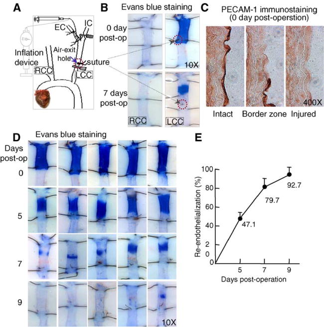
Fig. 1.

Mouse model of carotid artery air-dry endothelial denudation and reendothelialization. A. Schematic illustration of air-dry endothelial denudation. The LCC was dilated with 2 atmospheres of pressure, followed by endothelial denudation via injecting high-speed air through the carotid. A suture was placed to mark the air-exit hole. B. Evans blue staining. Animals were perfused with Evans blue at the indicated time. Excised carotids were pinned on dissecting plates. At the site of endothelial denudation, the vessel takes up dye and appears blue. C. PECAM-1 immunostaining. Acetone-fixed frozen longitudinal sections of completely or partially deendothelialized carotid arteries were immunostained with PECAM-1 antibody. The intact endothelium stained dark brown. The border zone of the injury site reveals the transition from absence to presence of endothelium. The injured section shows an absence of endothelium. D and E. Time course of post-injury reendothelialization. Images are representative of Evans blue stained mouse carotids harvested at the indicated time points. The area of endothelial denudation is defined as the branching point of EC, from IC to the suture marker at the air-exit hole. The graph shows the percentage of reendothelialization over time in the injured carotid. LCC, left common carotid; RCC, right common carotid; EC, external carotid; IC, internal carotid; PECAM-1, Platelet-Endothelial Cell Adhesion Molecule-1.
