
Figure 2. CMA is upregulated in response to double strand DNA damage.
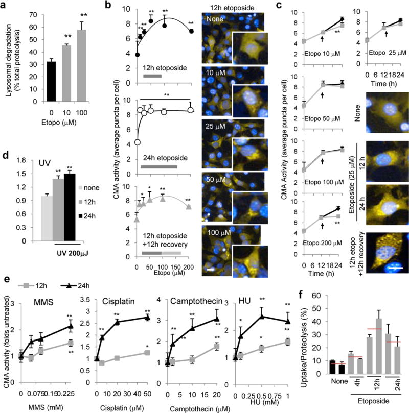
a. Lysosomal protein degradation in mouse fibroblasts treated with etoposide for 24h, n=6 wells in 3 independent experiments. b,c. CMA activity in cells expressing a photoactivatable CMA reporter and treated with increasing concentrations of etoposide for the indicated times (b) or for different times at the indicated concentrations of etoposide (c). Arrow indicates etoposide removal after 12h of treatment (grey lane) Right: Representative images of the indicated conditions. Inserts in b show higher magnification images. n=3 independent experiments, >200 cells/experiment were counted. d,e. CMA activity measured as in b at the indicated times after UV exposure (d) or at 12 or 24h after the indicated treatments (e). n=3 independent experiments, >200 cells/experiment. f. CMA activity against a pool of radiolabeled cytosolic proteins of lysosomes isolated from livers of mice untreated (none) or at different times after a single etoposide injection. n=3 independent experiments and the means of two animals per group are shown with average value (red line). All values are mean+s.e.m except panel F that are mean and range. (unpaired two-tailed t-test). *P<0.005 or ***P<0.0005. Scale bar: 10μm.