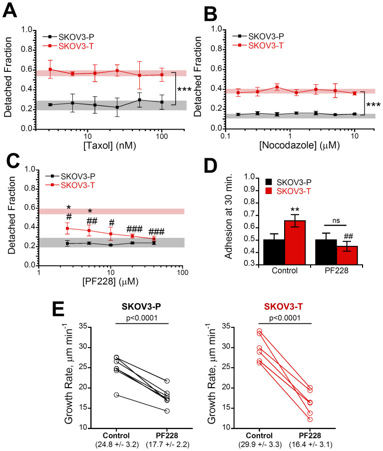
Figure 5. Alterations in adhesion and microtubule dynamics in Taxol-resistant cells are reversible with FAK inhibition.
(A) SKOV3 parent (-P) and Taxol-resistant (-T) were pre-incubated with 1:1 serial dilutions ranging of Taxol ranging from 3.13–100 nM for 4 hours to stabilize microtubules before repeating the adhesion strength assay and displayed no change from the untreated controls (shaded region) (N = 3). (B) The adhesion strength assay was repeated using 4 hour nocodazole pre-treatment with 1:1 serial dilutions ranging from 0.16–10 μM to depolymerize microtubules (N = 3). In order to prevent changes from increased Rho activity upon nocodazole upon washout the experiment was carried out in the drug-containing media instead, producing a slightly higher baseline for SKOV3-T. (C) To inhibit focal adhesion signaling cells were pre-incubated with 1:1 serial dilutions of focal adhesion kinase inhibitor PF228 ranging from 2.5–40 μM for 4 hours before running the adhesion strength assay demonstrating selective recovery of adhesion force in Taxol-resistant cells relative to their untreated controls (red shaded region) to become equivalent with parent control cells (gray shaded region) (N = 3). (D) To determine the effects of FAK inhibition on attachment kinetics, cells were pre-incubated with 10 μM PF228 for 30 minutes in solution and allowed to adhere for 30 minutes revealing FAK inhibition decreased attachment kinetics in Taxol-resistant cells (N = 3). (E) Cells transfected with mCherry-EB3 were imaged and then treated with 10 μM PF228 for four hours before re-imaging revealing a significant (p < 0.0001) decrease in both parent and Taxol-resistant cells to a statistically equal value. Values listed in parenthesis are given as mean ± std. All other values given as mean ± SEM; significance is indicated relative to matched parent population with *′s and relative to solvent treated control #′s. *P < 0.05, **P < 0.01,***P < 0.001.

