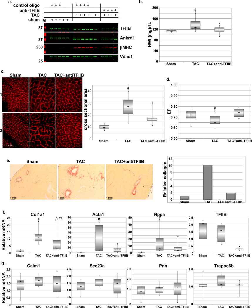
Figure 5. Acute antisense inhibition of TFIIB reduces cardiac hypertrophy-induced gene expression and the increase in heart weight.
Twelve-week old, male, mice were subjected to a sham or transverse aortic constriction (TAC) operation. After 1d the mice were randomly selected for injection with saline or 15 mg/Kg LNA-modified control or antisense TFIIB (anti-TFIIB) oligo, as indicated (n=7 each). a. After 3 weeks the hearts were isolated, protein was extracted and subjected to Western blotting for the specified genes. One experimental set is shown here; the second set is shown in the online data (set = same day surgery for all included mice). b. Box-plot of the heart weights of the mice, adjusted to tibial length. c. Similarly treated heart were isolated, sectioned and stained with wheat germ agglutinin for delineating cross sectional area. Panels 1 and 2 represent sections from 2 different hearts. Cross sectional area of 10 myocyte/section/heart was measured and plotted (graph, right). * is p < 0.05 v. sham, # is p < 0.5 v. TAC+anti-TFIIB. d. All mice were analyzed by echocardiography before sacrifice and the ejection fraction calculated and plotted. * is p < 0.05 v. sham, # is p < 0.5 v. TAC+anti-TFIIB. e. Sectioned hearts were stained with Sirius red for estimating collagen content (n=2). The red-stained collaged was quantified, averaged, and plotted (graph, right). f. Total mRNA was extracted from all hearts and select hypertrophy-related genes were quantified by qPCR. * is p < 0.05 v. sham, # is p < 0.5 v. TAC+anti-TFIIB. g. * is p < 0.05 v. sham, # is p < 0.5 v. TAC+anti-TFIIB. g. Total mRNA was extracted from all hearts and select housekeeping genes were quantified by qPCR. * is p < 0.05 v. sham, # is p < 0.5 v. TAC+anti-TFIIB. g. * is p < 0.05 v. sham, # is p < 0.5 v. TAC+anti-TFIIB.

