Figure 2.
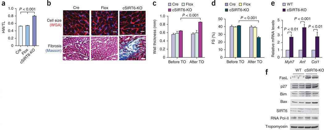
Cardiac-specific deletion of SIRT6 causes cardiac hypertrophy and fibrosis. (a) HW/TL ratio of control mice (Cre, α-MHC–Cre; Flox, SIRT6flox/flox) and SIRT6flox/flox–α-MHC–Cre mice injected with tamoxifen to generate a cardiac-specific SIRT6 deletion (cSIRT6-KO). Data are presented as the mean ± s.d. n = 9–15 mice per group. Student’s t test was used to calculate the P value. (b) Heart sections of cardiac-specific SIRT6 knockout and control mice stained with WGA to demarcate cell boundaries (top; scale bars, 10 µm) or with Masson’s trichrome to detect fibrosis (bottom; scale bars, 40 µm). (c,d) Left ventricular wall thickness and fractional shortening of control and cardiac-specific SIRT6 knockout mice before and after tamoxifen (TO) injection. n = 5 mice per group. (e) mRNA levels of the indicated genes in heart samples of control and cardiac-specific SIRT6 knockout mice. Data are presented as the mean ± s.d. n = 4 per group. (f) Western blots showing the expression of the indicated cell death markers in the same hearts as in e. For c–e, Student’s t test was used to calculate the P value.

