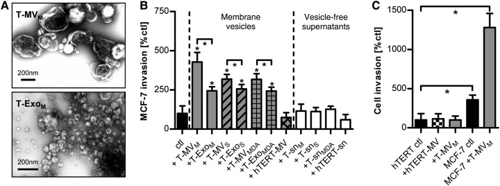
Figure 1.
T-EV induce breast cancer invasion. (A) Electron microscopy (TEM) of T-MV and T-Exo. (B) Microinvasion assay of MCF-7 breast cancer cells stimulated with T-MV, T-Exo, and MV from normal epithelial cells (hTERT-MV) (all at 1 µg/ml), as well as the respective particle-free supernatants (sn) (mean ± SD, n = 3, *P < 0.001). Suffix M: vesicles/supernatant from MCF-7 cells; S: SK-BR-3; MDA: MDA-MB231; hTERT: hTERT-HME1. (C) Comparative analysis of cell invasion of MCF-7 and benign hTERT-HME1 cells stimulated with MV (10 µg/ml) (mean ± SD, n = 3, *P < 0.001).

