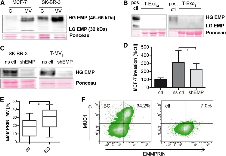
Figure 3.
EMMPRIN is expressed on T-MV in vitro and in vivo and contributes to their proinvasive phenotype. (A and B) Western blots showing the expression of highly (HG) and lowly glycosylated (LG) EMMPRIN (EMP) in whole cell lysates (C) and T-MV (A) as well as the corresponding T-Exo (B) of both breast cancer cell lines. (C) Western blots showing stable knockdown of EMMPRIN via shRNA (shEMP) in SK-BR-3 cells and MV (ns ctl, non-sense control). (D) Microinvasion assay of MCF-7 cells exposed to T-MVS (1 µg/ml) from EMMPRIN knockdown cells (mean ± SD, n = 3, *P < 0.01). (E) Total MV from peripheral blood of metastatic breast cancer (BC) patients and matched controls (ctl). The percentage of EMMPRIN-positive MV was quantified by flow cytometry. (F) Double staining for EMMPRIN and MUC1 of MV from a BC and a control patient (representative density plots).

