3.

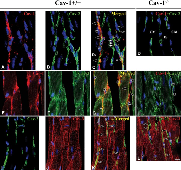
Co-localization between caveolins in Cav-1+/+ and Cav-1−/− mouse LVM. In Cav-1+/+ 3D reconstructed images (-45° rotation of y-axis) shows Cav-1 (red) localizes at both FB (nucleus: large asterisks) and CEC (nucleus: small asterisks), but Cav-2 (green) localizes only at CEC. Partial co-localization of Cav-1 and Cav-2 occurs at CEC (open arrows), but sometimes Cav-1 without Cav-2 occurs at CEC membrane (closed arrows) (A–C). Cav-1 (red) co-localizes well with Cav-3 (green) at CM plasma membrane (large open arrow), FB (nucleus: large asterisks) interconnecting with tails (small open arrow) and CEC (nucleus: small asterisks). Also, Cav-1 immunoreactivity occurs near CM Z-lines and partial co-localization with Cav-3 (E–G). Cav-2 (green) co-localizes partially with Cav-3 (red) at CEC (arrows). CEC make frequently anastomotic structure (asterisk) (I–K). In Cav-1−/− Cav-2 (green) immunore-activity occurs regardless of Cav-1 at CEC (d). Cav-3 (green), immunoreactivity occurs also at CM plasma membranes, Z-lines and FB (asterisks) (H). Co-localization (open arrows) of Cav-2 (green) and Cav-3 (red) at CEC is similar to that in Cav-1+/+(I). CM is cardiomyocyte and IS is interstitial space. Scale bar is 10 mm for all images.
