8.

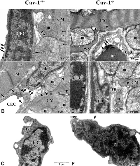
Electron micrographs of CM, FB and CEC in Cav-1+/+ and Cav-1−/−mouse LVM. In Cav-1+/+ mouse A–C shows that CM, FB and CEC are closely located. Some caveolae (Cav, arrows) reveal at CM sar-colemma and a number of (Cav, arrows) reveal at CEC membrane. Well-developed sarcoplasmic retic-ulums (SR, closed thin arrows) are continuously arranged between myofilaments in CM and a few endoplasmic reticulums (ER, open thin arrows) reveal near by Cav at CEC membrane. Sacs of T-tubules (arrowheads) reveal adjacent to Cav and SR of CM and large mito-chondria (m) locate adjacent to sar-colemma. FB does not have Cav but have well-developed rough ER. In Cav-1−/− mouse D–F shows that CM, FB and CEC are closely located as Cav-1+/+ mouse. A few Cav (arrow) reveal at the sarcolemma and some flat-shaped SR (closed thin arrows) along the sarcolemma are discontinuously located in CM. Sacs of T-tubules (arrowheads) reveal adjacent to SR. CEC have a few Cav. FB does not have Cav, but a few have it. FB-p, process of fibroblast.
