3.

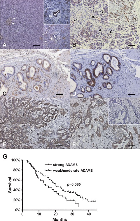
ADAM8 expression and localization in the normal pancreas, CP and PDAC tissues: Immunohistochemistry was performed as described in the ‘Materials and methods’ section. (A) ADAM8 expression in normal pancreatic islets (arrows) and acinar cells and ductal cells (lower inset). Consecutive section stained with CK19 confirming ductal cells (upper inset). (B) ADAM8 expression in CP tissues (arrows indicate tubular complexes). Consecutive section stained with CK19 confirming ductal cells/tubular complexes (inset). (C, D) ADAM8 (C) and CK19 (D) staining in pancreatic cancer cells of PDAC tissues. (E, F) Strong ADAM8 staining in pancreatic cancer cells of PDAC tissues. Note the absent staining in a consecutive negative control tissue section (inset). Horizontal lines represent the scale bar of 50 μm. (G) Survival curves of PDAC patients (n = 99) with weak/moderate ADAM8 staining versus strong ADAM8 staining (P= 0.065).
