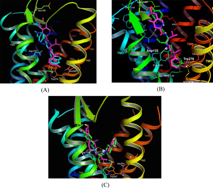
Figure 3.
(A) Binding model of JDTic in the NOP receptor crystal structure 4EA3. The TM helices are color-coded and annotated. The NOP antagonist C-24 bound in the NOP crystal structure is shown in cyan, whereas JDTic is in magenta. Key interacting amino acids are as indicated. (B) Binding orientation of opioid pan antagonist AT-076 in the NOP crystal structure. AT-076 (green) is superimposed on JDTic (magenta) in the NOP binding pocket. Both ligands bind in an extended conformation in the NOP receptor. (C) Binding model of AT-076 in the KOP receptor crystal structure 4DJH. AT-076 (green) is superimposed on the bound orientation of JDTic (magenta) in the KOP crystal structure. Note that AT-076 binds KOP in a similar V-shaped orientation as JDTic, but with the phenylpiperidine and isoquinoline moieties oriented exactly opposite to those of JDTic.

