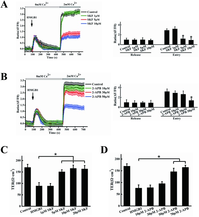
Fig 4. SKF96365 and 2-APB reduce Ca2+ influx and HMGB1-induced permeability.
EA.hy926 cells were preincubated with 1, 5, 10 μM SKF96365 (A), or 10, 30, 50 μM 2-APB(B) or vehicle (DMSO), then stimulated with 200 ng/ml HMGB1, followed by the addition of 2 mM CaCl2. Intracellular calcium transients were measured using an Olympus FV1000 confocal microscope. Peak intracellular Ca2+ was quantified during intracellular release or extracellular Ca2+ influx. EA.hy926 cells were plated in the upper part of transwell chambers until the formation of a tight monolayer. The cells were preincubated with 1, 5, 10, 20 μM SKF96365 (C), or 10, 30, 50, 70 μM 2-APB (D) for 1 h, respectively. HMGB1 200 ng/ml was then added and the cells were incubated for an additional 24 h. After incubation, the integrity of the tight junctions was assessed by measuring the TER. Data are presented as mean ± SD of three independent experiments. *Indicates significant difference compared with the control group (P<0.05).

