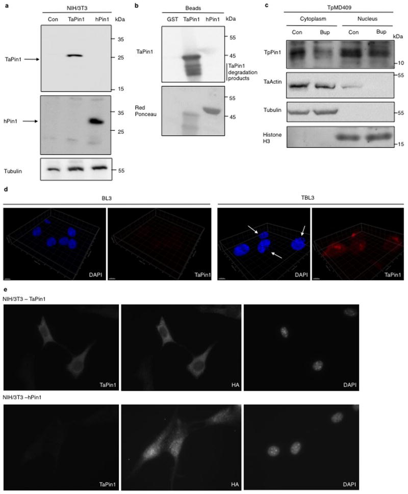
Extended Data Fig. 4. Characterization of the TaPin1 antibody and secretion of TpPin1 in Theileria parva.
a The antibody raised against TaPin1 specifically recognizes Theileria version in TaPin1-stably transfected NIH3T3 cells but not the empty vector in Control-stably transfected NIH3T3 cells or the human version in hPin1-stably transfected NIH3T3 cells. Both Theileria and Human versions of Pin1 are present respectively in TaPin1-stably transfected NIH3T3 and hPin1-stably transfected NIH3T3 cells.
b. The antibody raised against TaPin1 specifically recognizes GST-TaPin1 but not GST and GST-hPin1 beads.
c. TpPin1 protein was detected in the nuclear and cytoplasmic fractions of T.parva-infected TpMD409 cells and decreased upon Buparvaquone (Bup) treatment. “Con” indicates Control. Antibodies recognizing apicomplexan actin, bovine Tubulin or bovine Histone H3 were used as controls.
d. TaPin1 expression in BL3 or parasite-infected TBL3 cells was examined by confocal microscopy analysis using an antibody raised specifically against Theileria TaPin1, counterstaining with DAPI. 3D reconstruction in BL3 and TBL3 cells are shown.
e. Immunostaining of TaPin1 and HA in NIH-3T3 stably expressing TaPin1 and NIH-3T3 stably expressing hPin1 counterstaining with DAPI.
All the results are representatives of 3 independent experiments.

