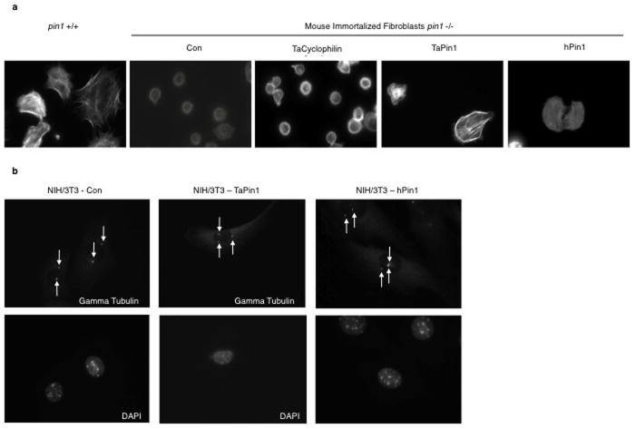
Extended Data Fig. 5. TaPin1 functionally replaces hPin1.
a. The HA-tagged version of TaPin1 and hPin1 can rescue cell spreading defects in knockout pin1-/- murine immortalized fibroblats. Cell spreading was assessed by Phalloïdin-TRITC staining. “Con” indicates Control = transfection with the appropriate empty vector.
b. TaPin1 causes centrosome amplification. NIH3T3 fibroblasts stably expressing TaPin1 or hPin1 were arrested at the G1/S transition by aphidicolin, stained with anti-γ-tubulin antibody (arrow) and counterstained with DAPI.
These photos are representative of cell in 3 independent experiments

