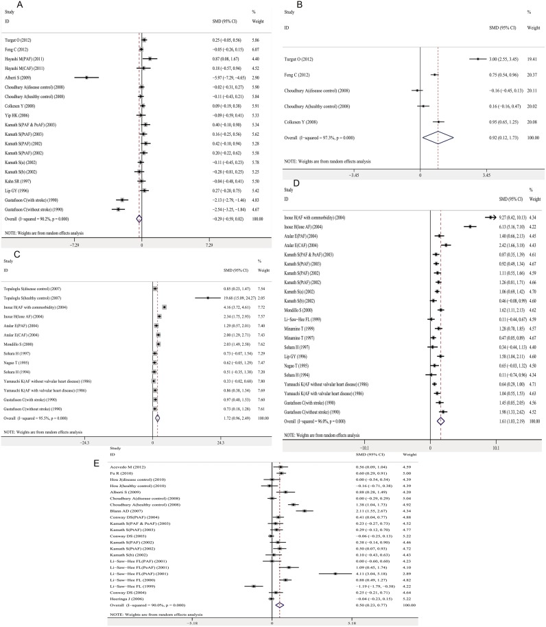
Fig 2. Association between platelet activation markers and AF.
A. platelet count and AF; B. Mean platelet volume and AF; C. Platelet factor-4 and AF; D. β-thromboglobulin and AF; E. P-selectin and AF. Forest plots of SMD and overall SMD with 95% CI between AF cases and controls. Black diamonds indicate the SMD, with the size of the square inversely proportional to its variance, and horizontal lines represent the 95% CI. The pooled results are indicated by the black hollow diamond. AF, atrial fibrillation; MPV, mean platelet volume; PF-4, platelet factor-4; BTG, β-thromboglobulin; PAF, paroxysmal AF; PeAF, persistent AF; PtAF, permanent AF; CAF, chronic AF; SMD, standardized mean difference.

