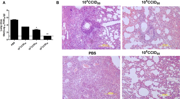
Fig 6. Lung viral titers and histopathological changes of vaccinated mice following challenge with live influenza Ah01/H7N9 virus.
Two weeks after the boost, vaccinated mice were i.n. infected with Ah01/H7N9 (50LD50) and lung tissues were collected 3 days later. (A) Viral titers in the lung of infected mice. The data were determined in triplicate by CCID50 assay in MDCK cells 3 days post infection and expressed as log10 CCID50/g tissue. Data are means ± SD. *p<0.01; ** p<0.001 (B) Lung histopathological changes following virus challenge. Representative histopathological images of lung damage by H&E staining from five mice per group.

