Figure 2.
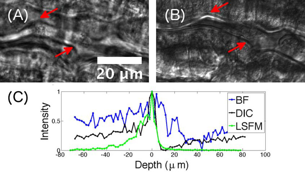
Characterization of various imaging techniques. (A) Brightfield and (B) DICM images of the same region of the intestine of a 7 dpf (days post fertilization) larval zebrafish. In the DICM image, features such as the gut edge (arrows) are clearly evident. (C) Pixel intensity, normalized to a peak value of 1, as a function of depth relative to the focal plane from images of a fluorescent polystyrene microsphere embedded in agar imaged with BF (brightfield), DICM, and LSFM, providing a measure of the depth of focus, and optical sectioning ability, of each method.

