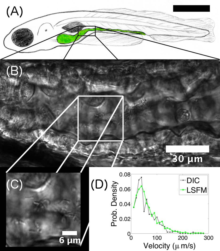
Figure 3.
(A) Schematic illustration of a larval zebrafish, with the intestine highlighted in green. The scale bar is approximately 0.5 mm. (B) DICM image of a section of the intestine of a 6 dpf zebrafish. The gut boundaries are clearly evident. (C) Part of the image in (B), showing individual rod-like Vibrio cholerae bacteria. (D) Velocity distribution of Vibrio cholerae in the gut, obtained from either LSFM or DICM imaging.

