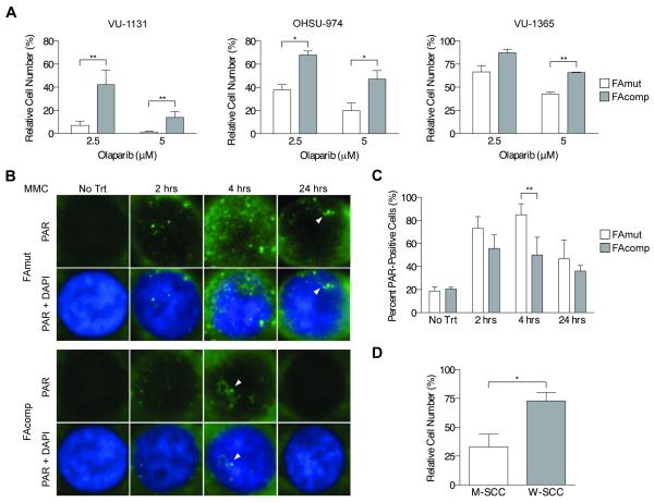
Figure 6. PARP inhibitor sensitivity of human and murine FA HNSCC cells.
A) Cellular growth assays on isogenic human HNSCC cells exposed to the PARP1/PARP2 inhibitor olaparib shows uniform sensitivity of FAmut cell lines. * p < 0.05; ** p < 0.01 (t-test). B) Immunofluorescence for PAR foci in VU-1131 cells shows increased PAR foci formation in FAmut cells over a 24-hour course of MMC treatment. Results shown are representative of two (no treatment) or three (2, 4, and 24 hours) independent experiments of each time point, each with similar results. C) Quantification of intranuclear PAR foci in VU-1131 cells over a 24-hour course of MMC treatment reveals a significantly increased number of PAR foci in FAmut cells at 4 hours of exposure. ** p < 0.01 (t-test). D) Cellular growth assays performed on murine Fancc−/− (M-SCC) and WT (W-SCC) HNSCC cell lines treated with olaparib. shows significantly increased sensitivity of Fancc−/− cell lines. * p < 0.05.

