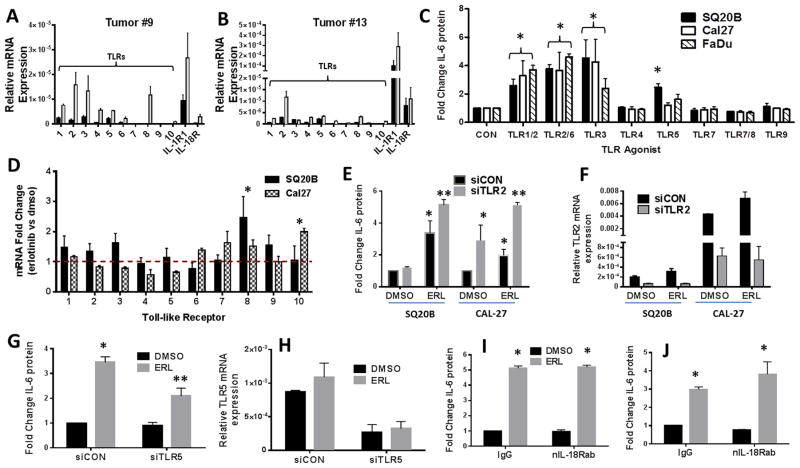
Figure 4. Role of TLR signaling in erlotinib-induced IL-6 in HNSCC cells.
A,B: RNA isolated from two HNSCC tumors (#9 (A) and #13 (B)) (gray bars) and matched normal tissue (black bars) was analyzed for TLR1-10, IL-1R and IL-18R gene expression by RTPCR. C: SQ20B, Cal27 and FaDu cells were treated with TLR agonists as described in the Methods section. Secreted IL-6 was measured by ELISA. D: SQ20B and Cal-27 were treated with DMSO or 5μM erlotinib (ERL) for 48 hours. Cells were analyzed by RTPCR for expression of TLR genes. Values were normalized to 18S mRNA levels, and reported as fold change over DMSO (set at 1, dotted line). E–H: SQ20B or Cal-27 cells were transfected with scrambled siRNA control (siCON), siRNA targeted against TLR2 (siTLR2) (E,F), or siRNA targeted against TLR5 (siTLR5) (G,H), treated with DMSO or 5μM ERL, then analyzed for IL-6. Knockdown of respective TLRs were confirmed by RTPCR (F,H). SQ20B (I) and Cal27 (J) cells were treated with IgG or an IL-18R neutralizing antibody (nIL-18Rab, 0.5 ug/mL) for two hours prior to DMSO or ERL (5μM) before IL-6 analysis. N=3, errors bars = standard error of the mean (SEM). *p<0.05 versus control; **p<0.05 versus ERL.

